


Start - Aktualności - Help in the treatment of chronic wounds
Help in the treatment of chronic wounds
Treating chronic wounds is complex, time-consuming and often expensive. Without appropriate therapy, these wounds will not heal spontaneously - this especially applies to diabetic wounds or venous ulcers. The treatment process can be supported by a wound scanner, which is being developed as part of the Woundscanning project with the participation of scientists from the Faculty of Biomedical Engineering of the Silesian University of Technology.
The scanner is used to image and diagnose difficult-to-heal wounds. Scanning is performed in a non-contact manner, which makes the procedure sterile, hygienic and does not cause pain to the patient. The construction of the device is carried out by a company from the Yoshi Group - WoundScanning sp. z o. o. and is the implementation of research results conducted by the Department of Medical Informatics and Artificial Intelligence in the field of imaging and analysis of chronic wounds. The Silesian University of Technology is a subcontractor in the project, carrying out conceptual work in the field of scanner construction and in the processing and analysis of collected data. The university is also responsible for developing appropriate software.
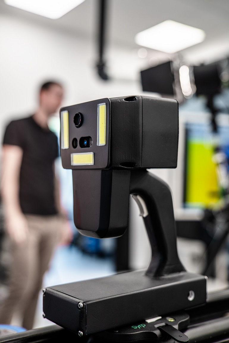
The scanner allows to take a 3D image of the wound using a ToF (Time-of-Flight) camera. – The device is equipped with a camera and a thermal imaging camera. It is designed and calibrated in such a way that it is possible to superimpose both a photographic and thermal image on a three-dimensional surface model. Wound scanning using 3D ToF-based technology brings many benefits, including high precision, speed of measurement, painlessness - says dr Jan Juszczyk from the Department of Medical Informatics and Artificial Intelligence.
Previous research on the possibility of visualizing difficult-to-heal wounds by creating 3D models has demonstrated the usefulness of such visualization in monitoring the treatment process. – The scanner construction project is a further stage of this work. The goal is to develop a device that will allow a quick scan of the wound surface in a chronic wound treatment clinic, explains the scientist.
This scanning device will be used to document chronic and difficult-to-heal wounds, i.e. those that last longer than 6 weeks. These include postoperative wounds, diabetic foot, bedsores, burns, ulcers, and oncological lesions. The process of their treatment is often long and difficult to assess. One of the basic methods of verifying the progress of therapy is to compare the size of the wound surface between subsequent visits to the clinic. For this purpose, photographic documentation is made.
– On the one hand, the wound scanner will simplify this process, and on the other hand, it will allow for greater objectivity by providing a 3D model of the wound and enriching it with the information contained in the thermal image. Obtaining reliable and possibly accurate imaging data translates into a greater amount of valuable information for the doctor - explains dr Juszczyk.
The scanner is a compact device with a housing resembling typical hand-held 3D scanners. It consists of a module containing sensors and a viewing screen connected to the handle. The device is designed to be operated with one hand, even when wearing gloves, it is powered by batteries and connects to the computer wirelessly, so it does not restrict the operator's freedom of movement.
– Performing a scan is very simple. Just point the device towards the wound so that the entire wound is visible on the preview screen and press the button. The rest of the registration happens automatically. The scanner is a non-contact device and its operation is similar to operating a simple camera - explains the scientist.
Importantly, the wound scanner does not use any radiation that is harmful to the patient.
– The main benefit for the patient is faster and more precise determination of effective therapy. Thanks to this, if the expected treatment results are not achieved, the doctor will be able to react earlier by modifying or changing the therapy - emphasizes the scientist.
In turn, for medical staff, the greatest benefit will be the shortening and improvement of the process of creating photographic documentation and ensuring more precise measurement results.
Work on the wound scanner is nearing completion. A prototype has already been created. Now scientists from the Faculty of Biomedical Engineering of the Silesian University of Technology are testing the software.