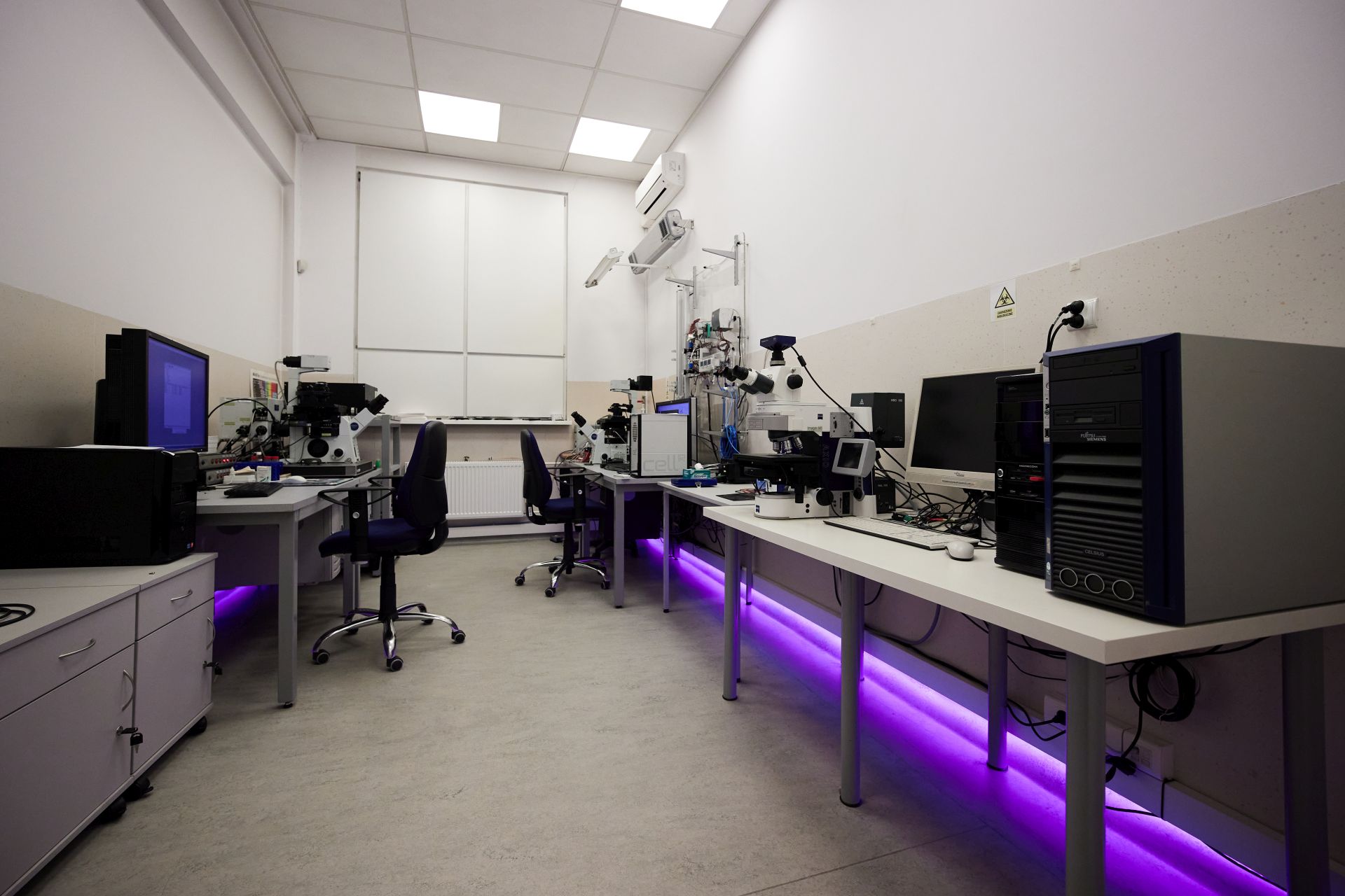
In the Section of Microscopic Research and Visualization, a set of microscopes enable advanced research in molecular biology, microbiology, cytology, histology, and chemistry.
In the section we have the following systems:
An Olympus FluoView FV1000 confocal scanning microscope with a spectral detection system with a unique SIM scanner is an inverted microscope that allows you to visualize and analyze rapid changes taking place in biological samples. Thanks to its high resolution and efficiency, the system allows for multidimensional imaging (3D with time dimension) of cell structures and tissue morphology, including the analysis of molecular location. The confocal technique based on scanning lasers provides the best quality images while maintaining low phototoxicity. The system is equipped with an incubation chamber for live-cell studies, which, combined with a unique system of simultaneous stimulation and observation of the object, and high quality and contrast of the images obtained allows for microscopic observations, 3D reconstruction, as well as the analysis in 4D of rapid structural changes taking place in living organisms.
Our system and software allow us to conduct experiments with the use of advanced techniques, i.e .:
- Laser light stimulation of biological processes and photoconversion
- FRAP - Fluorescence Recovery After Photobleaching )
- FLIP - Fluorescence Loss In Photobleaching )
- FLIM -I Fluorescence Lifetime Imaging Microscopy )
- Assembling confocal images for large biological objects
- Analysis of changes in the position and mobility of molecules - quantitative analysis of colocalization
The IX81 motorized inverted microscope with Cell ^ R imaging system with a high-quality Hamamatsu digital camera, live-cell chamber, and high-speed motorized stage allows for qualitative and quantitative fully automated analysis of intracellular processes for large populations of cells or tissue preparations. The minimal cytotoxicity in the observations allows for the survival analysis of processes, i.e. protein co-localization and expression, the cell cycle, the analysis of damage to cell structures, or processes related to cell proliferation and death.
Fluorescence microscopes by Zeiss and Olympus with a computer imaging system allow for the quantitative analysis of images for various types of samples. Advanced software for automatic image analysis allows you to speed up the performed statistical analyses.
Lab Supervisor:
dr hab. inż. prof. Pol. Śl. Sebastian Student, tel: 237 2906, Sebastian.Student@polsl.pl